
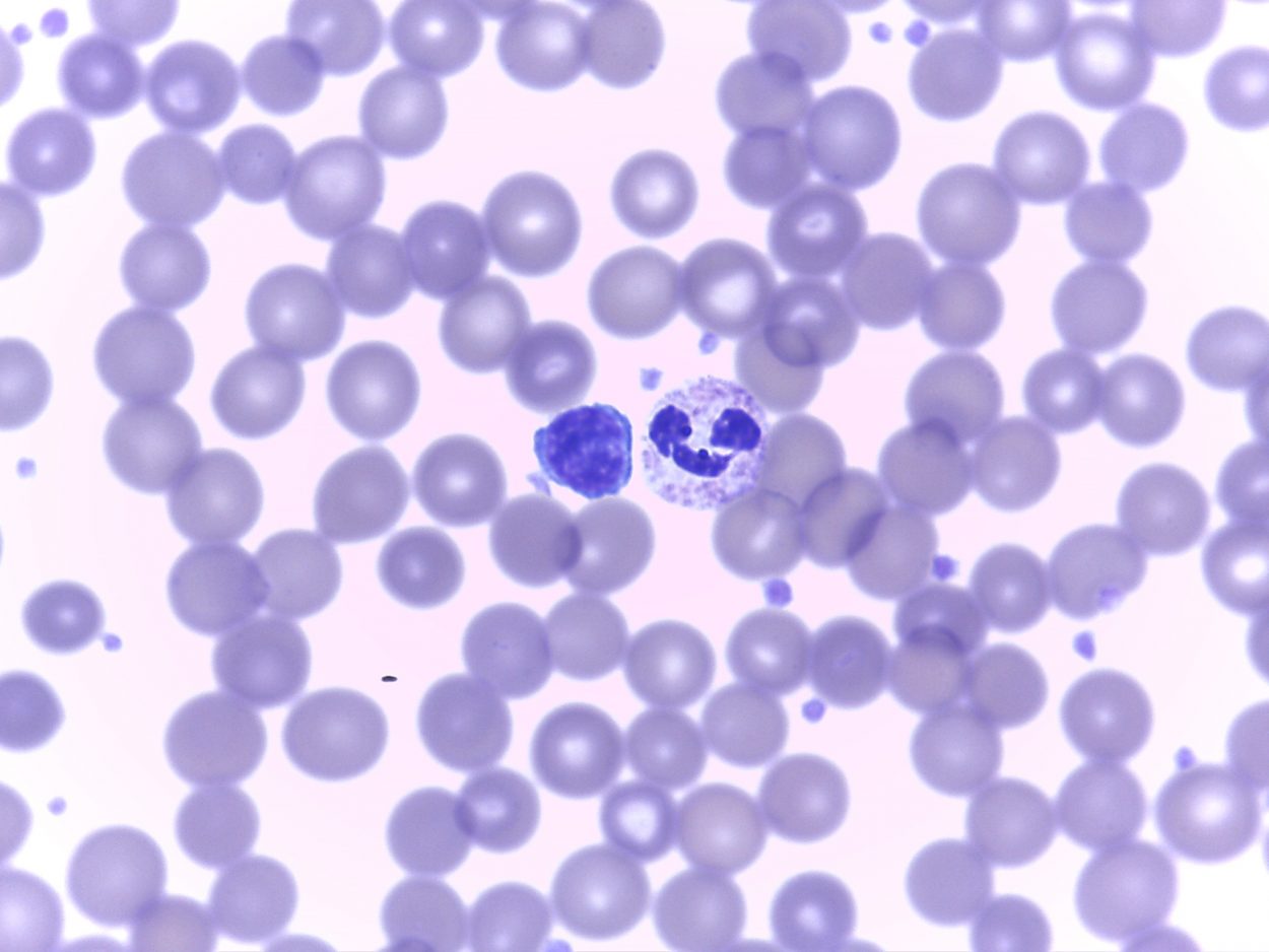
A blood smear specimen at the Petersburg lab shows lymph and mature netrophil. Photo courtesy of Liz Bacom
The laboratory is abuzz with electronics. Computers and other equipment fill the open room.
This is where things like blood and urine get tested, specimens that one might expect to be tested in a hospital laboratory. But there are other kinds of testing going on here that just might surprise you.
Liz Bacom is the Lab and Imaging Manager.
“We test potable water, cannery water, and pool water for chloroforms,” Bacom says. “We’re looking for an indicator organism that e-coli and if that’s present then that indicates that the water’s been contaminated with warm blooded fecal materials.”
Bacom says the lab is certified with Alaska’s Department of Environmental Conservation.
“So, we do the borough, we do residences, if you live out the road and you want to test your water, you can bring a sample into us,” Bacom says.
The medical center’s lab workers are all considered generalists so they can each work in chemistry, hematology, and microbiology. Bacom says having tests done in house can speed up treatment for patients.
“The benefit of having a good microbiology department is a number of reasons,” Bacom says. “First of all, if you’ve got a urine culture, a urine infection and you want to find out if you need to be treated or not we can usually tell within the next day whether there is something there.”
The more unusual tests still get sent out three times a week to Seattle to be conducted there.
Next to the microbiology room is the hematology and chemistry lab. This is where about 600 samples of blood get tested during the medical center’s health fair every other Spring.
Lori Miller is one of the hospital’s six medical technologists. She’s sitting in a lab coat in front of a large computer screen looking at what looks like hundreds of circles.
“We’re looking at a blood smear for one of our patients,” Miller says. “These are red blood cells, all these little dots. And these are actually different types of white blood cells. This is a monocyte and this is a lymphocyte.”
The image is a mystery to me, but to her, it’s like reading a kind of blood map.
“If you look at the red blood cells as well as part of our differential you can tell if the patient is anemic by what the red blood cells look like,” Miller says. “These all look pretty good because they’re nice and red. We can tell a lot just from one slide of blood.”
Lab testing is only one department. There’s also the imaging department. That’s found down the hallway in separate rooms for CTs, X-Rays, Ultrasounds, and Mamographies. Two staff people work there.
All the images are taken in these rooms and sent out to a radiologist group in Eugene, Oregon. They are on-call 24 hours a day, seven days a week. It’s all digital and results can be returned as quick as twenty minutes.
“When I first started working here it was wet chemistry,” Bacom says, “so we’d take a picture, an x-ray, put it into a tank in a dark room and develop it, it come out in 90 seconds, stick it up on the view box and the doctor here would take a look at it and go, ‘well, not sure if it’s a C spine fracture or not, we’re going to have to ship the patient out and so what this has done is really reduce the number of medivacs.”
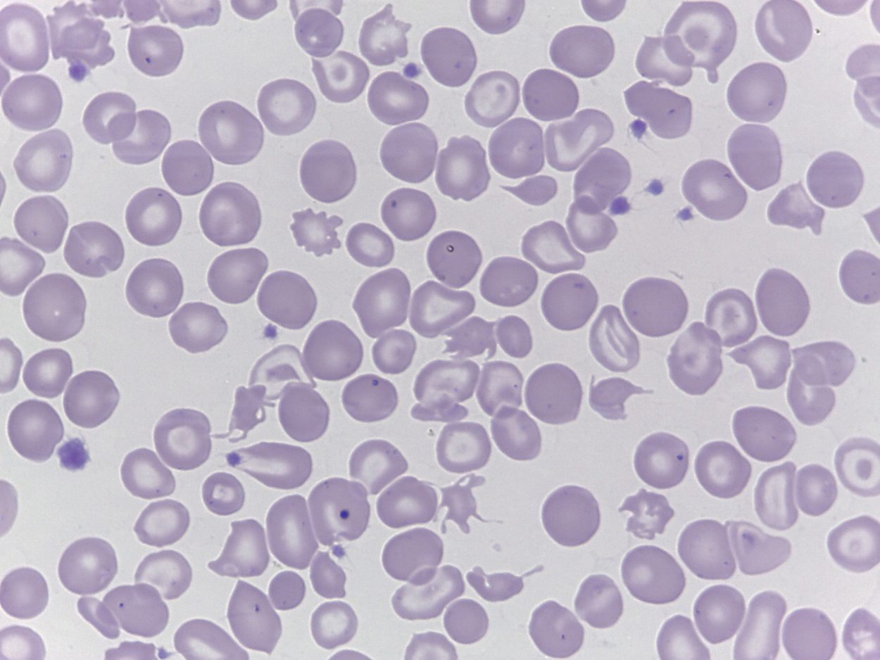
A blood smear at the Petersburg lab shows anemia. Photo courtesy of Liz Bacom
The images are digital and can be shared. The lab works with a pathologist in Bellingham, Washington who can help them remotely trouble shoot any questionable images.
The C T Scanner is a 16-slice machine which means it’s not the top of the line but not the bottom either. It can’t look at details in the heart for example, but it can help patients who had a stroke.
“One of the things we have to find out is whether it’s a bleed in the head and whether or not it’s a bleed will determine how that patient’s going to be treated and you have to have that information really quick because the sooner you start therapy, the better the outcome for the patient,” Bacom says.
The CT machine gives physicians a good idea if further testing is needed.
For basic imaging needs, say for a broken bone, the X ray machine in the next room does the trick. It is about 15 years old and the parts aren’t available anymore. It’s still used regularly and Bacom hopes for funding to replace it.
Like the X-ray machine, the mammography machine is also older. It’s still affective for checking abnormalities in breasts. . . it’s just not digital.
“This is the only non-digital mammography unit in the state of Alaska,” Bacom says. “That means that the quality of the images is good but it’s not as sensitive as a pure digital image. So what we have to do is take an image and process it through a separate computer system that enhances it so it is as close to digital as it can be without being completely digital.”
The mammography machine is used about 15 to 20 times a month. The machine is worth over $200,000 and Bacom says the money is just not there to replace it now.
Then there’s the portable ultrasound machine which uses sound waves to look inside the body. This one was upgraded last year.
And to maintain all of this imaging equipment? It takes specialists. They fly into Petersburg once a year to check on the equipment to make sure it’s running like it should.


